The cranial or anterior cruciate ligament is one of the 4 main ligaments stabilizing the knee or stifle joint. Most commonly, rupture of the cranial cruciate ligament is due to degenerative changes and chronic inflammation that is sometimes compounded by trauma.
Approximately 50% of dogs with complete tears also have a tear in the medial meniscus, which is a cartilage pad on top of the tibia or shin bone. Since cranial cruciate tears are a result of progressive, degenerative changes, often times the sudden onset of non-weight bearing lameness is proceeded by intermittent, chronic bouts of variable lameness.
Rupture of the cranial cruciate is such a common cause for sudden onset of hind limb lameness in the dog that any sudden onset of hind limb lameness is due to cruciate disease unless proven otherwise. Generally the pain associated with a torn cruciate ligament is such that there is little to no weight bearing on the affected leg. If the cruciate tear is not treated then there likely will be some improvement seen over several weeks, but lameness will persist and the resultant arthritis will be worse with no surgical treatment.
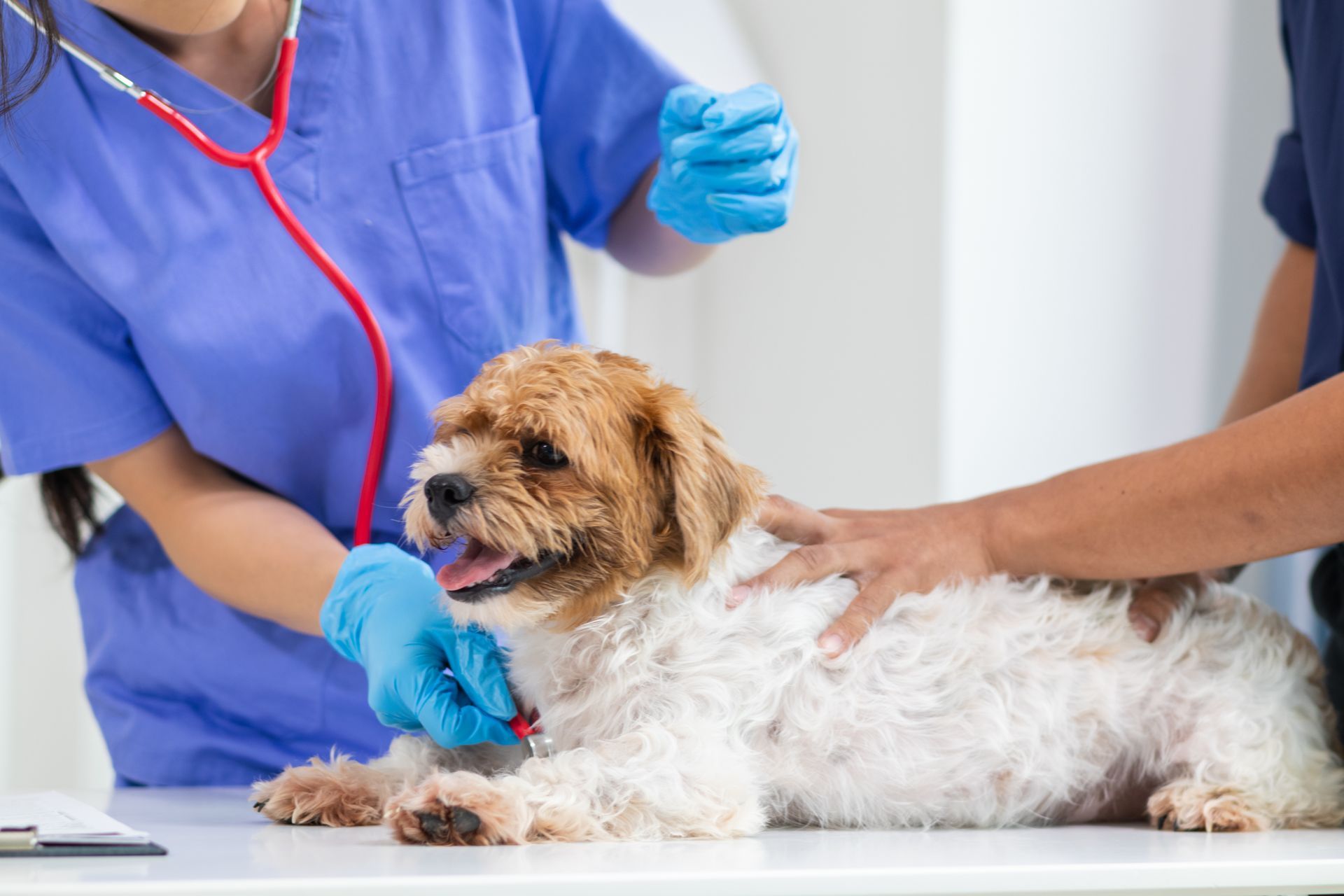
Diagnosis of cruciate disease can typically be made based on physical examination and x-rays of the stifle along with possibly other joints on the same leg to eliminate other problems contributing to the lameness. Typically, physical exam demonstrates a “drawer” sign with cruciate ligament rupture. This is a movement of the top of the tibia (shin bone) forward from underneath the femur (thigh bone) that would not be possible if the cruciate ligament was intact. It is possible with dogs, especially large dogs, for them to tense muscles so tightly during examination that they must be sedated to accurately do this test. Also a partial rupture may not result in an obvious “drawer” sign.
X-rays are helpful for a number of reasons. While the cruciate ligament itself cannot be visualized with x-rays, a characteristic pattern of effusion (change in normal x-ray density) occurs with cruciate disease. It is also valuable to know the extent of arthritis, changes of which can be seen on x-rays, to prognosticate on extent of recovery of pain free movement after surgery.
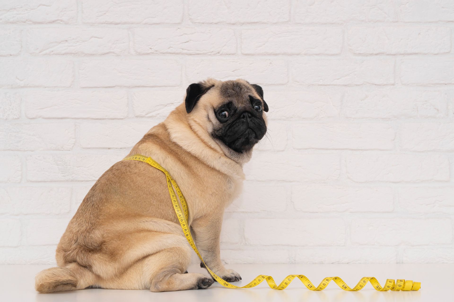
Rupture of the cranial cruciate ligament is most commonly due to an idiopathic degenerative condition within the stifle joint. Many large breeds of dogs are particularly at risk and a study in Labradors has found a genetic risk. For a Labrador that ruptures a cranial cruciate ligament about 62% of the risk is genetic. A genetic test is available to determine risk in Labradors. The test accurately predicts at 98% if a Labrador will rupture a cruciate ligament. A recently published study indicated that neutering before 12 months of age is associated with greater risk of cruciate rupture in Labradors. Weight control is important for decreasing load and stress on the stifle joint. In one study, obesity quadrupled the risk of cranial cruciate ligament rupture.
Treatment for a ruptured cruciate ligament is surgery. For dogs weighing more than 30 pounds, less than 20% have an acceptable outcome without surgical treatment. There are 2 primary objectives with surgery. The first is to accurately identify and remove any torn meniscus. This is the cartilage pad on top of the tibia (shin bone) that is attached to the cranial cruciate ligament and is often torn when a cranial cruciate ligament tears. If the torn meniscus is left, it will result in persistent pain like a pebble under your foot in a shoe.
The cruciate ligament’s function is to prevent the movement of the tibia forward when pressure is directed downwards on it by the femur. The second objective of surgery is to fix what a torn cruciate ligament is unable to do. Several procedures have been developed. The generally preferred procedure for active dogs and dogs over 45 pounds is the tibial plateau leveling osteotomy (TPLO). This procedure essentially changes the angle or slope of the top of the tibia so that when force is directed downwards through the femur, the pressure tries to push the tibia backwards which it cannot do because of other ligaments. This provides dynamic stabilization of the stifle.
With rupture of the cranial cruciate ligament there will be development of some arthritis. In order to minimize this, the rupture must be treated surgically and in a timely manner. Following surgery, weight reduction if indicated along with chondroprotective agents can be helpful. For some, medical therapy with a non-steroidal anti-inflammatory and other pain relieving medication maybe needed. Most importantly, with cranial cruciate ligament rupture in order to prevent debilitating arthritis, if not already present, one needs timely surgical intervention.